Darkfield Microscopy (DF) and Hyperspectral Imaging (HSI)

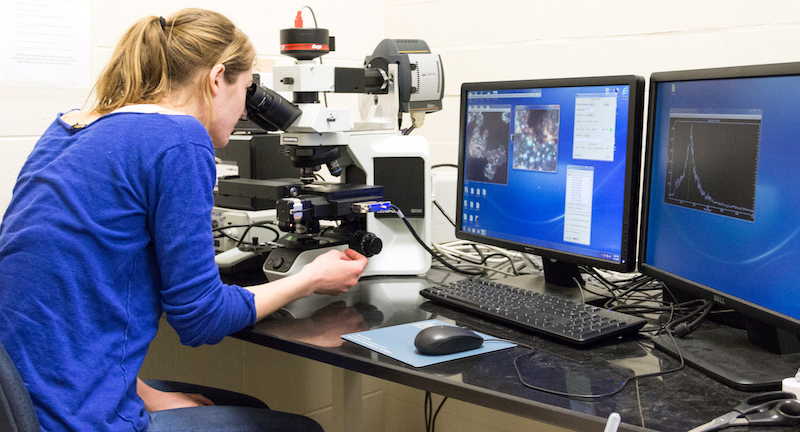
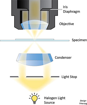
 Darkfield (DF) microscopy coupled to hyperspectral imaging (HSI) yields a detailed map of surface plasmon resonance (SPR) spectra for each pixel. The SPR frequency of nanoparticles (NPs) depend on particle size, shape, dielectric properties, aggregate morphology, surface modification and refractive index of the surrounding medium. In general, the SPR absorption peak red shifts to longer wavelengths with increase in particle size. The recording of an absorbance spectrum for each pixel in the image is element dependent (i.e. an elemental fingerprint) and therefore, it is possible to identify the presence of defined NPs, which allows to generate a distribution map of NPs and their aggregates in the specimen. Visualization of NPs at the LM level including HSI has the great advantage that a high number of cells or a larger surface area can be investigated in a short time (hours to days) avoiding the time-consuming embedding and section procedure that one would have to apply for TEM (weeks to months). It also allows to investigate real-time effects by tracking the same NPs in the same sample over time.
This content has been updated on 12 May 2022 at 16 h 36 min.
